Upper Thigh Anatomy / Mri Of The Thigh Radiology Key - This image added by admin.. The upper leg is often called the thigh. The hamstrings are those three muscles that are located in the back of the thighs. Anatomically, it is part of the lower limb. There are five muscles in the anterior thigh compartment: Learn more about the hardest working muscle in the body with this quick guide to the anatomy of the heart.
They work closely with your quadriceps muscles at the front of your thigh, your gluteal muscles, and your calf muscles to ensure proper movement of your leg and hip. Teachme anatomy part of the teachme series the medical information on this site is provided as an information resource only, and is not to be used or relied on for any diagnostic or treatment purposes. Muscles are named according to their shape, location, or a combination. Charlotte o'leary bsc, mbchb • reviewer: A deep, shooting pain in the upper leg can also be caused by deep vein thrombosis, spinal stenosis, or a thigh bone infection.
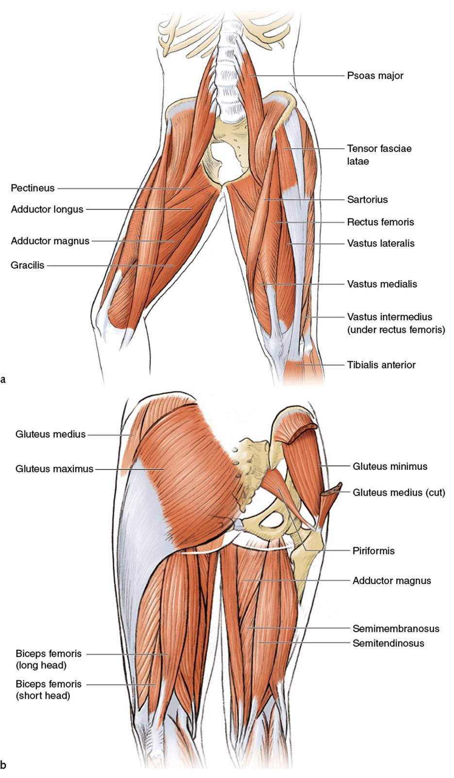
Upper Legs Running Anatomy Sports Anatomy from doctorlib.info The upper leg is often called the thigh. This webpage presents the anatomical structures found on thigh mri. Anterior muscles extend your legs and flex your thighs. They work closely with your quadriceps muscles at the front of your thigh, your gluteal muscles, and your calf muscles to ensure proper movement of your leg and hip. You can click the image to magnify if you cannot see clearly. The largest of the quadriceps muscles, it's located on the outside of the thigh and runs from the top of your femur (thighbone) down to your kneecap (patella). Sartorius, and the four quadriceps muscles; The musculature of the thigh can be split into three sections;
The hamstrings are those three muscles that are located in the back of the thighs.
•medial thigh muscles•adductor longus muscle•adductor magnus muscle•adductor. Work the small muscles of your inner thighs to find ease in all sorts. A deep, shooting pain in the upper leg can also be caused by deep vein thrombosis, spinal stenosis, or a thigh bone infection. They work closely with your quadriceps muscles at the front of your thigh, your gluteal muscles, and your calf muscles to ensure proper movement of your leg and hip. The musculature of the thigh can be split into three sections; Tools:wacom intuos3 tabletadobe photoshopcamtasia studiomusic by mefor free art resources, like photoshop br. Also called the thigh bone, this is the longest bone in the body.it. We think this is the most useful anatomy picture that you need. Related posts of muscle anatomy of upper thigh human muscle anatomy. Ebraheim's educational animated video describes muscle anatomy of the thigh. The thigh bone, or femur, is the large upper leg bone that connects the lower leg bones (knee joint) to the pelvic bone (hip joint). Pretty self explanatory from the title i think. The muscles that make up the quadriceps are the strongest and leanest of all muscles in the body.
This mri brain cross sectional anatomy tool is absolutely free to use. You can click the image to magnify if you cannot see clearly. The upper leg, in particular, is comprised of bones and muscles that are susceptible to injury, particularly when excess strain is placed upon them. Human muscle anatomy 12 photos of the human muscle anatomy human anatomy muscle questions, human anatomy muscles clay learning system, human muscle anatomy head, human muscle anatomy leg, human muscle anatomy worksheet, human muscles, human anatomy muscle questions, human anatomy muscles clay learning system, human muscle. The posterior upper leg muscles provide your knees with mobility (extension, flexion and rotation) and strength.
Ligaments Tendons And Muscles Of The Hip Joint Naples Best Hip Surgeon from zehrcenter.com This image added by admin. The hamstrings are those three muscles that are located in the back of the thighs. Meanwhile, the vastus lateralis is on the side of the thigh, while the vastus intermedius is hidden below the rectus femoris(5). Human muscle anatomy 12 photos of the human muscle anatomy human anatomy muscle questions, human anatomy muscles clay learning system, human muscle anatomy head, human muscle anatomy leg, human muscle anatomy worksheet, human muscles, human anatomy muscle questions, human anatomy muscles clay learning system, human muscle. At the top, there is the pelvis bones which do not belong to the lower it starts from the outer surface of the ilium bone of the pelvis. This mri brain cross sectional anatomy tool is absolutely free to use. The thigh muscles don't just move your legs. Tools:wacom intuos3 tabletadobe photoshopcamtasia studiomusic by mefor free art resources, like photoshop br.
The muscles that make up the quadriceps are the strongest and leanest of all muscles in the body.
The upper legs consist of three main muscles: Legs give us the freedom to run, walk, jump, climb, and negotiate the world around us. February 25, 2021 reading time: Upper thigh anatomy (page 1). At the top, there is the pelvis bones which do not belong to the lower it starts from the outer surface of the ilium bone of the pelvis. Atlas of body sections, ct and mri images, fourth edition. 2, vastus medialis & intermedius muscles. Anatomynote.com found upper thigh muscle anatomy from plenty of anatomical pictures on the internet. You can click the image to magnify if you cannot see clearly. The rectus femoris is located in the center of the thigh, while the vastus medialis is in the middle of the said body part. It's the area that runs from the hip to the knee in each leg. Benjamin ma, md, professor, chief, sports medicine and shoulder service, ucsf department of orthopaedic surgery, san francisco, ca. This image added by admin.
Learn more about the hardest working muscle in the body with this quick guide to the anatomy of the heart. Each compartment has a distinct innervation and function. Spicermanyt at checkout for 40% off this tutorial! Benjamin ma, md, professor, chief, sports medicine and shoulder service, ucsf department of orthopaedic surgery, san francisco, ca. Upper inner thigh anatomy :
Anterior Compartment Of Thigh Muscles Their Action And Nerve Supply Anatomy Qa from i1.wp.com Fasciae of the hip and thigh author: They work closely with your quadriceps muscles at the front of your thigh, your gluteal muscles, and your calf muscles to ensure proper movement of your leg and hip. The four muscles all extend the lower leg. Also called the thigh bone, this is the longest bone in the body.it. The upper leg is often called the thigh. Anatomynote.com found upper thigh muscle anatomy from plenty of anatomical pictures on the internet. Upper thigh cross sectional anatomy : We think this is the most useful anatomy picture that you need.
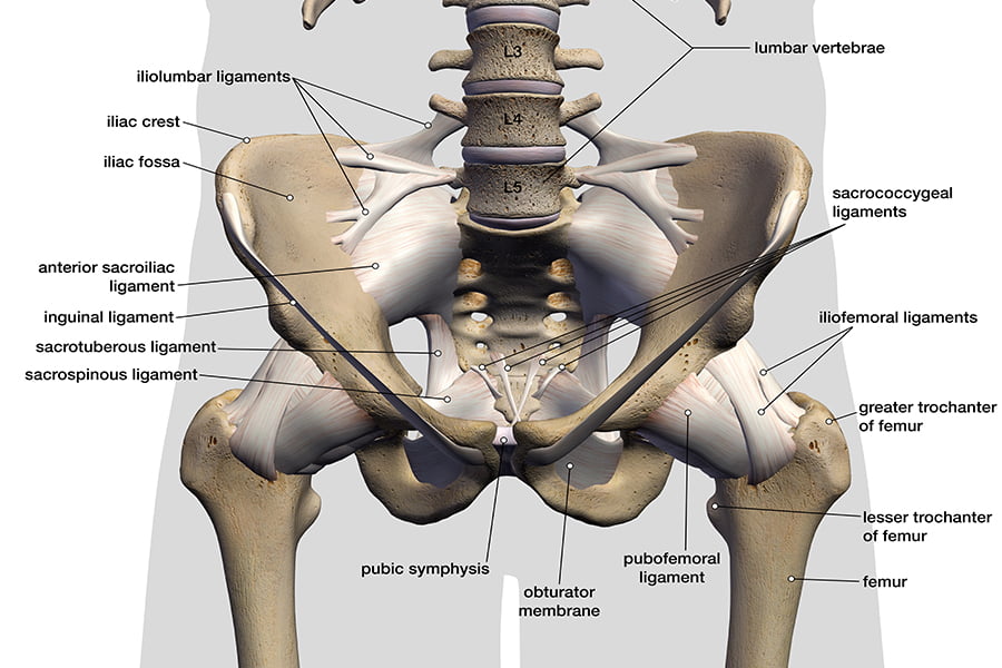
Muscles and ligaments work together to support the spine, hold it upright, and control movement during rest and activity.
The muscles located within the posterior compartment of the thigh are the biceps femoris, semitendinosus and semimembranosus. The four muscles all extend the lower leg. Like the adductors, the abductors are also responsible for stabilizing your knees during athletic and everyday movement. They have a lot to do with how your hips move. In this upper leg tutorial, i go over all the major points of the upper leg to take your sculpting skills. 8 minutes fascia is a band of connective tissue located beneath the skin, which encloses and separates muscles.there are two main types of fascia: They work closely with your quadriceps muscles at the front of your thigh, your gluteal muscles, and your calf muscles to ensure proper movement of your leg and hip. February 25, 2021 reading time: These muscles help us to allow the. The rectus femoris is located in the center of the thigh, while the vastus medialis is in the middle of the said body part. Ebraheim's educational animated video describes muscle anatomy of the thigh. Spicermanyt at checkout for 40% off this tutorial! The largest of the quadriceps muscles, it's located on the outside of the thigh and runs from the top of your femur (thighbone) down to your kneecap (patella).